Dental Disease In Pets, The Silent Killer – Part 2
![]() Rayya The Vet
Rayya The Vet ![]() Apr 14, 2013
Apr 14, 2013 ![]() Highlight, NGOs, Rayya The Vet
Highlight, NGOs, Rayya The Vet ![]() 0 comments
0 comments
| Tweet |
Now to pick up where I left off in my last post; allow me to highlight the most common symptoms your pets may present with when suffering from a tooth ache and this will include some nauseating pictures.
In dogs, the subtle signs they may display include:
- Favoring soft food,
- Burying their raw bone instead of tackling it head on (few exceptions are those dogs that enjoy hiding their bone until it ripens) and/or preferentially chewing on one side of their mouth (this can easily be missed without very close observation).
- A uniform build-up of plaque on one side of your dog’s mouth will often indicate preferential chewing on the unaffected side and this can be diagnosed by your veterinarian during a routine health check.
One of the main reasons that your dog may prefer to chew on one side of his/her mouth is if they have fractured a tooth.
As in the case of Buster below, his owner actually heard him yelp while chewing on his bone. It did not stop him from eating but he was obviously struggling to chomp his food. Examination revealed he had fractured his upper right 4th premolar (carnassial) tooth. We proceeded with a full general anesthetic and extraction of the affected tooth.


As for Lucy, this jack russell terrier came in for a dental check and was not showing any obvious signs of discomfort to her owners. I incidentally discovered she had uniform buildup of plaque affecting only the left side of her mouth.
It turned out she had a slab fracture to her upper left 4th premolar tooth. I removed the fragmented surface of the tooth and did not proceed with a full extraction. My reasoning was the underlying tooth did not appear to have any exposed pulp. Her owners are fully aware she may require a full extraction later on.


Tinker was another dog that did not display overt discomfort when eating. He had a mild bad breath but he would not allow us to examine his mouth. We proceeded with a full general anesthetic and discovered he had a decaying upper right 4th premolar (carnassial) tooth decay. I extracted this tooth as my dental probe revealed a large pocket of loose gums.
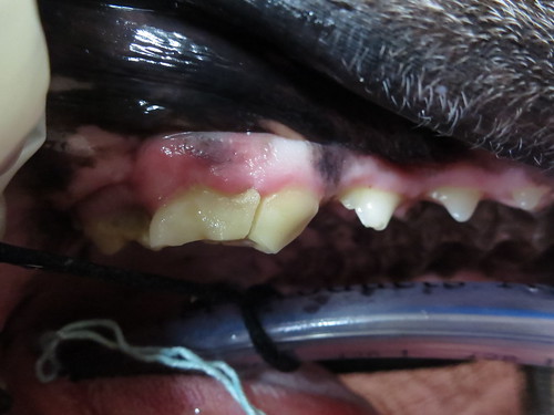
One of the most typical reasons dogs are brought to the vets for an oral examination is the development of intolerable bad breath referred to as halitosis. The bad breath develops in association with inflamed or ulcerated gingiva and mostly due to the excessive buildup of tartar and plaque.
Buzz is a rescued cocker spaniel that required two dental procedures only a few months apart. In the first procedure, he was castrated and had a dental scale. It was noted that he had have severe gingival recession affecting most of his teeth. Only a few months later, he developed recurrent halitosis and painful gums and so we proceeded with another dental. This time around, we extracted the most severely affected teeth. He may still require further extractions in the future.
Please note that the yellowish staining observed over his upper left canine should be in fact covered with gum tissue.

Here, you can clearly observe the exposure of the left roots of his lower first and second premolars. This illustrates the severity of his gingival recession. You should not be seeing the roots of your dog’s teeth.

Here are some cases with severe plaque buildup:
Molly is a greyhound prone to the development of plaque especially on her upper canines. Regrettably, she is head shy and will not allow her owner to brush her teeth. She will very likely require 6 monthly to annual dental scales to prevent progression of this condition.

This was not Meggie’s first dental procedure but was a much needed one. The level of plaque buildup over her upper right 3rd and 4th premolars is horrendous.

Sophie is a 6 year old Italian grey hound with a severely infected upper right 4th premolar tooth. This was her second dental procedure. Poor little one had already lost a decent set of teeth. If her dental problems were addressed earlier, we may have been able to rescue many of her teeth and not just resort to multiple dental extractions.

Badger is a very old Dachshund cross with some serious plaque buildup affecting most of his teeth. I had been trying to convince his owners to perform a dental for almost one year. They were petrified of losing him under general anesthetic since he was already diagnosed and being treated for heart disease.
He recovered brilliantly from the procedure and is still maintained on his heart tablets.
His owners were so thankful that I convinced them to go ahead as they have noted a huge boost in his spirit and livelihood since this was done.
Can you even begin to imagine the pain he was enduring with a mouth like this ? Believe it or not, he was still eating without any obvious discomfort!?!

So for those of you that still truly rely on the fact that your dog’s appetite is a good indicator on how well your dog is coping with their dental disease, please think again.
Now let me share with you the most classic presentation of dogs requiring immediate dental attention. This is what your dog could look like when things go pear shaped with his/her tooth.
Often clients do not realise the lump is associated with dental disease.
Your dog can acutely develop a tooth root abscess and the swelling in front of the eye is the point of drainage for the infection.
Check out poor Holden, a young dog that developed a tooth root abscess.

Unfortunately, this may be a situation you can’t always prevent in your pet as the root of the upper premolar or molar may have rotted with or without any obvious external damage to the visible tooth. In some canine patients, we can easily determine the culprit tooth.
Having said that, sometimes it requires a lot of probing and flushing to definitively determine which tooth requires extraction.
This is another case of a tooth root abscess. Toby was a very difficult patient to handle and required a full general anesthetic to allow us to examine his mouth. We then proceeded with the curative treatment which involves the extraction of the offender tooth. A course of antibiotics may have resolved the swelling but it would have only been a temporary solution.
In this picture, I was using an alligator forceps to determine which tooth was the source of his festering infection. I had already extracted one loose upper molar but discovered it was not the one contributing to the abscess.

It is crucial to probe the discharging sinus to definitively confirm which tooth requires extraction. There is nothing worse than removing a loose tooth and assuming it is the one contributing to the abscess and then finding out you were wrong. Your patient will have to come back for another general anesthetic because you failed to extract all the necessary teeth.
Another very interesting dental case that I dealt with was Gabby. Her owner had brought her into the clinic on several occasions as she was concerned about recurrent sneezing episodes that partially responded to repeat courses of antibiotics. Her oral examination did not reveal any obvious dental disease.
I decided it was about time we put her under general anesthetic to look up her nose and do further workup.
I quickly discovered the source of her problem when my dental probe disappeared into her gums. You can now understand how easy it was to misdiagnose her condition. Her canines appear healthy and have very little tartar buildup and no visible gingival recession to indicate significant dental disease.

Her upper canines were quite rotten and I extracted both of them without any difficulty. She recovered well and guess what, she stopped sneezing! A note to veterinarians out there, Dachshunds seem quite predisposed to this condition. So please add upper canine tooth root infection to your list of differentials for the next sneezing Dachshund you are presented with.
This case illustrates the importance of conducting a general anesthetic to appropriately assess certain dental cases.
Not many canines or felines (for that matter) will allow you to use a dental probe to inspect their teeth while conscious.
This picture is just to illustrate the depth of the root of a dog’s canine. Boof had an gingival swelling revolving around his lower right canine which prompted my colleague to extract it and biopsy the abnormal gingiva.

This poor dog was noise phobic and in the process of escaping from the backyard, he managed to pull out his upper left canine. It was merely attached by some gingival tissue. Now that would have hurt A LOT!

Now it is quite important that I mention oral lumps as these can be another reason you book your dog in for a dental check. In general, oral lumps are often bad news. However, some oral lumps are related to dental disease. You may not be able to visualize some of the oral lumps as they are tucked way back and hiding behind a large tooth.
If your dog’s breath suddenly goes really bad, then make sure to get him/her immediately checked out by your local veterinarian as he/she may have developed an oral lump.
In this case, the owner quickly identified her dog’s issue and brought her in for a check. Holly had developed a fleshy lump around her upper right most lateral incisor.

We proceeded with a full general anesthetic and upon closer inspection, it appeared like it was reactive gingival tissue surrounding an infected tooth. We cracked the plaque that was over the infected incisor and thoroughly scaled it. We collected a biopsy sample of the affected gum tissue to rule out an aggressive oral tumor.
Thankfully the biopsy results confirmed my suspicion of it just being a reactive benign epulis.
This is another case that was booked in for a dental and we incidentally discovered the patient had reactive gum tissue associated with severe dental disease.
The picture is taken after a thorough dental scale was conducted.

As for this case,we incidentally discovered multiple oral lumps that may or may not be associated with dental disease. Some of the lumps were in close proximity with infected teeth while others were not.
We recommended collecting multiple biopsies as we were concerned they may be nasty oral tumors.
Unfortunately, the owner could not afford sending the lumps away or further treatment. I’m crossing my fingers that they were only associated with plaque and have not progressed since the dental was done.


Last but not least, here are some photographs of pre and post dental procedures I have conducted.
I am always pleased with the finished product and hope more owners will realise the importance of dental hygiene in their dogs. We should be tackling your dog’s dental health early on and not leave it until the dental only involves multiple extractions.
The cases below are listed in order of severity (starting from least to most):
Ernie pre dental:

Ernie post dental:

Tuppy pre dental:

Tuppy post dental:

Boof pre dental:

Boof post dental:

Lilly pre dental:


Lilly post dental:


Gracie pre dental:

A close up of Gracie’s premolars.

Gracie post dental:

Monkey pre dental:

Monkey post dental:

Gidgette pre dental:

Gidgette post dental:

Star pre dental:


Frodo pre dental:


Lilly pre dental


Star, Frodo and Lilly don’t have any post dental photographs.
Why you may ask? Well they barely had any teeth left after the procedure.
Please do your best to prevent your dogs from reaching these dogs’ state of dental disease.
I think I have pumped you with enough information for one session. I will discuss feline dentistry in the next post so make sure to come back and read the last dental sequel.
And before I forget, for those of you that live near my clinic, please take advantage of our 25% discount on all dental procedures (dogs and cats) this month! If you happen to live close by, please book in your pet for a free dental check to see if your pet could do with a dental.
Related articles
- Dental Disease in Pets, the Silent Killer – Part 1 (rayyathevet.com)
Source: http://rayyathevet.com/2013/04/14/dental-disease-in-pets-the..
| Tweet |
Facebook Comments