Oh No,I Discovered A Lump On My Pet! Part 1
![]() Rayya The Vet
Rayya The Vet ![]() Jan 14, 2013
Jan 14, 2013 ![]() Highlight, NGOs, Rayya The Vet
Highlight, NGOs, Rayya The Vet ![]() 0 comments
0 comments
| Tweet |
It must be very scary to discover a lump on your pet especially if it a decent size. I ask you not to panic and I hope this post will guide you on what to do next. I will be including lots of pictures and some may not be so pretty.
The rule of thumb with lumps is if it is growing too quickly and bothering your pet (appears red or inflamed) then it needs to be attended to immediately. However, this does not apply to all lumps. Some skin tumors can be slow growing but may still have a potential to spread to other areas. Bottom line, if you notice a lump on your pet, then get it checked out by your local veterinarian, better be safe than sorry!
Your veterinarian should always offer to do a fine needle aspirate to identify the lump as palpation alone is not diagnostic.
Some lumps don’t aspirate well and so a diagnosis can’t be made without collecting a biopsy sample. I routinely do fine needle aspirates on lumps and if I can’t identify the cells under the microscope, then I recommend sending off the sample to the pathologist or collecting a biopsy sample. Otherwise, you have the choice of measuring the lump and keeping a very close eye on it and it if starts to grow quickly then book in your pet for surgical removal of the lump.
Lipomas
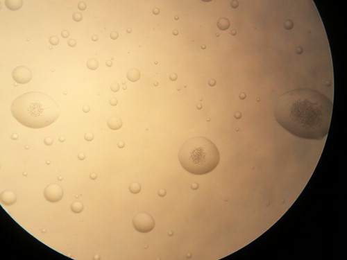
In geriatric patients, fatty lumps or ‘lipomas’ are quite common. It is an abnormal deposit of fat under the skin and we aren’t exactly sure of what instigates their development. Overweight patients and certain breeds like Labradors, Golden retrievers and Corgis seem to be quite prone to developing this type of lump. They are usually soft and unattached to the underlying tissue and benign. Lipomas are easily diagnosed with a fine needle aspirate.
This is a lipoma examined under the microscope. It appears like water droplets on a slide.

These lumps must be closely monitored as they can occasionally become cancerous. We don’t often resect these lumps as they generally recur. However, if they are causing discomfort for our patients or impeding their mobility due to their location or size e.g. in the axillary region (constant friction), then we recommend removing them. We also surgically remove them if they grow too big as they can burst. Occasionally, they can grow between muscles and feel quite hard and be mistaken for nasty lumps.
The take home massage is just because ‘lipomas’ are common in geriatrics, it does not mean we must assume it is one without getting it checked and that includes a fine needle aspirate being performed.
One of my most adorable geriatric patients came in as his owner noticed a sudden growth over his shoulder. It was not painful but had grown very rapidly and was quite large; about the size of a soccer ball. Palpation revealed it was a very hard lump and the fine needle aspirate was inconclusive. We proceeded with immediate surgery to investigate the lump.
Oscar the 10 year labrador

Oscar under general anesthetic investigating his very large hard lump.

This is an inside view of Oscar’s lump. At that stage, I was still very concerned and collected a biopsy sample and sent it off to the pathologist. Thankfully it turned out to be a lipoma.

Histiocyte
Another very common lump that usually affects young dogs is a ‘histiocytoma’ or ‘histiocyte’. This is an allergic type of benign tumor that mostly occurs in younger dogs. They take the form of raised fur-less reddened lumps. On gross examination, they are indistinct to ‘mast cells’ which are very nasty tumors
It is important to confirm they are histiocytes and that would involve sending either a fine needle aspirate or biopsy sample to a pathologist to examine. Generally they spontaneously resolve after a few weeks.
I tend to prescribe antihistamine for patients with this type of lump as it will help reduce the irritation and itching associated with it.
‘Simba’ is a 2 year old golden retriever that developed a 5 cent piece reddened fur-less lump under his jaw. It was itchy and that’s how the owner noticed it. We collected fine needle aspirates and sent them away to the pathologist and results came back confirming it is very likely a histiocyte. The lump disappeared on its own.

This young jack russell terrier presented to me with this lump under its right eye and it was at a very tricky spot if surgical resection was required. I proceeded with a full general anesthetic and collected a biopsy sample of the lump. I needed a definitive diagnosis of the lump and the fine needle aspirates would not have been sufficient. Thankfully results confirmed it was a histiocyte and it also disappeared on its own.

‘Penny’ was only 6 months old and she suddenly developed a reddened fur-less lump over her right shoulder that kept growing rapidly until it reached the size of a lemon. We proceeded with surgical resection of the lump and sent off a biopsy sample from it which confirmed it was a histiocyte. This is a picture of her after she had recovered from her surgery. The scar is the war wound she was left with after her surgery.

Mast cell
Now I would like to talk about the dreaded ‘mast cell’ as this is one of the most common cancerous skin lumps in dogs. In my experience, these tumors can come in all shapes, sizes and forms. They can be irritating and sometimes they can appear quite inactive. I don’t think this tumor fulfills its typical textbook description. I always offer to do fine needle aspirates on any lumps discovered on your dog or cat to rule in/out a mast cell. I often diagnose mast cells on fine needle aspirates. In any case, the next step after that would be resection of the lump with a decent margin (minimum of 1.5-2 cm) and sending it off to the pathologist. For those mast cells in very difficult locations like distal limbs or high grade ones, I give the owners the option of taking their pet to a specialist for complete resection.
The specialists have the advantage of advanced imaging like CT which will delineate the margins of the mast cell and if it has already spread.
‘Ella’ is a 6 year old female desexed fox terrier cross in premium health. She presented to me for a swelling over her right elbow that would spontaneously shrink then bulge.


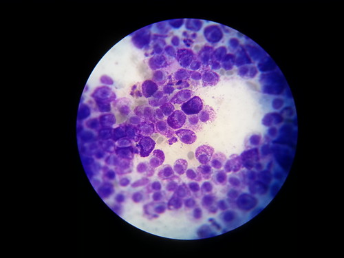
My examination of the affected area made me suspect she had a joint issue. Xrays were absolutely normal so I proceeded to collect a fine needle aspirate of her lump and this is a picture of the sample under the microscope. I was actually shocked to discover it was a mast cell as it didn’t fit the typical presentation.
In the pictures below, the densely purple cells with purple granules are the mast cells.

As for ‘Charlie’, he sure has been unlucky in that he has had 3 mast cells resected from him over the past 2-3 years. His first two mast cells were discovered when he was 5 years old. Thankfully his owners attended to them fairly quickly and they were all completely removed.

This is ‘Cindy’ and she is a 13 years old golden retriever that developed this very large lump fairly quickly behind her ear. My colleague proceeded with removing the lump and unfortunately an ear ablation was required for complete excision of the lump.

In cats, mast cells present in a much more diffuse manner and can be wide-spread throughout the skin of the patient. This is a picture of a geriatric cat with a very aggressive form of the mast cell tumor.

Bottom line is you should never judge a lump by the way it looks. Palpation alone is not sufficient to make a diagnosis. Your veterinarian should offer to do a fine needle aspirate for any routine lump check.
If the fine needle aspirate isn’t conclusive, then you must either closely monitor the lump or organize for it to be biopsied by your veterinarian.
I have discussed a handful of the most common skin lumps and have many more to go through. Please make sure to read the sequel to this post. And if you have any questions about the three lumps I mentioned in this blog, I’m all ears…
Source: http://rayyathevet.com/2013/01/14/oh-noi-discovered-a-lump-o..
| Tweet |
Facebook Comments